
All Images
Research News
Vaults: From Biological Mystery to Nanotech Workhorse?
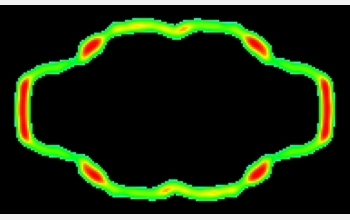
This image shows a typical cross-section through a vault particle, as derived from many electron micrographs of vaults in mouse cells. The highest protein density is shown in red, with lower densities in shades of green. The particle is 72.5 nanometers tall by 41 nanometers wide. The hollow interior is evident--and is easily big enough to enclose more familiar cellular structures, such as subunits of the ribosome.
Credit: Reprinted from Journal of Molecular Biology, Vol 344, 2004, Mikyas et al, "Cryoelectron Microscopy Imaging of Recombinant and Tissue Derived Vaults..." pp 91-105, with permission from Elsevier.
Download the high-resolution JPG version of the image. (68 KB)
Use your mouse to right-click (Mac users may need to Ctrl-click) the link above and choose the option that will save the file or target to your computer.
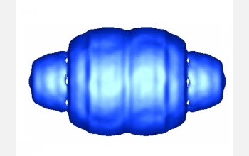
This image shows the overall structure of a vault particle, as derived from many electron micrographs of vaults in mouse cells. The particle is 72.5 nanometers tall by 41 nanometers wide.
Credit: Reprinted from Journal of Molecular Biology, Vol 344, 2004, Mikyas et al, "Cryoelectron Microscopy Imaging of Recombinant and Tissue Derived Vaults..." pp 91-105, with permission from Elsevier.
Download the high-resolution JPG version of the image. (115 KB)
Use your mouse to right-click (Mac users may need to Ctrl-click) the link above and choose the option that will save the file or target to your computer.
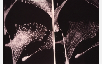
The image on the left shows a fibroblast cell that has been stained for vaults, while the image on the right shows the same cell after it has been stained for actin filaments. Actin filaments serve as cables and struts that help the cell move and control its shape; as such they are essential components of the cell's "cytoskeleton." A comparison between the two images shows that the vault particles tend to congregate along the filaments, especially at the tips--although no one knows why.
Credit: Nancy L. Kedersha and Leonard H. Rome, UCLA
Download the high-resolution JPG version of the image. (947 KB)
Use your mouse to right-click (Mac users may need to Ctrl-click) the link above and choose the option that will save the file or target to your computer.