
All Images
Research News
Researcher Says Life Evolved Between the Mica Sheets
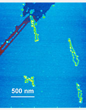
Biological molecules tend to bind well to mica. This atomic force microscope (AFM) image shows two yellow molecules on a blue mica surface, with a damaged purple-red area on the right where some of the top (blue) layer of mica peeled off.
Credit: Helen Greenwood Hansma, UC Santa Barbara
Download the high-resolution JPG version of the image. (473 KB)
Use your mouse to right-click (Mac users may need to Ctrl-click) the link above and choose the option that will save the file or target to your computer.
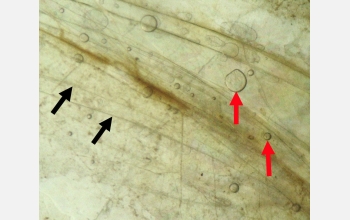
Photo of mica from an abandoned mica mine, with water between some layers, showing edges of mica sheets (black arrows), air bubbles in the water (red arrows) and brown bands of organic crud and dirt.
Credit: Helen Greenwood Hansma, UC Santa Barbara
Download the high-resolution JPG version of the image. (738 KB)
Use your mouse to right-click (Mac users may need to Ctrl-click) the link above and choose the option that will save the file or target to your computer.

Helen Greenwood Hansma, who became a grandmother during all this, poses with her grandchild.
Credit: Scott Hansma
Download the high-resolution JPG version of the image. (44 KB)
Use your mouse to right-click (Mac users may need to Ctrl-click) the link above and choose the option that will save the file or target to your computer.