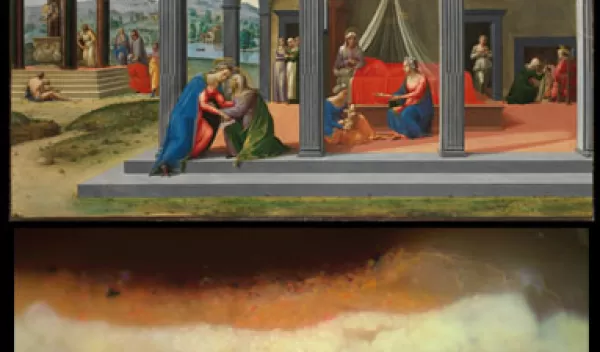
What Lies Beneath? Understanding Art Using Science
Proteins, enzymes, antibodies--when we hear these words, we are likely to conjure images in our heads of colorful molecular models, cancer, flu shots or even skin care. However, we seldom associate these terms with art. What does a protein, like collagen, for example, have to do with a Renaissance painting? The answer may surprise you.
At the Metropolitan Museum of Art (MMA) in New York City, in collaboration with Columbia University, and with funding through the National Science Foundation (NSF) Chemistry and Materials Research in Cultural Heritage Science program, scientists are employing their knowledge of both molecules and cutting-edge research techniques to uncover the material world of art--the organic compounds mixed with inorganic materials that compose what we see in a painting, a sculpture or even costumes.
Artworks are made of a diverse range of naturally occurring and synthetic materials, proteins being just one of those components. Knowing how a work of art is constructed is integral to understanding its historical significance, preservation or authenticity.
Whether a painting was made with egg tempera, as opposed to oil paint, may guide a conservator's approach to preserving a work, and inform a curator's interpretation.
Science offers the means of acquiring specific and relevant information about the materials used in a work of art. Scientists use a range of instrumental techniques to identify and study the ways in which these materials age and interact with their environment.
Organic compounds such as oils, resins, waxes, gums and animal-based protein binders, or glues, can be detected using Fourier-transform infrared spectroscopy (FTIR), and gas chromatography/mass spectrometry (GC/MS).
Both of those methods are staple tools for scientists in museums; however, the tools are not without their limitations. FTIR provides a fast means of determining the general class of material present in the sample. It is a helpful starting point, but it does not provide the specificity needed to characterize the compounds further. For example, an FTIR spectrum of a sample containing animal-based glue will indicate the presence of protein, but no information on the kind of protein.
GC/MS, on the other hand, gives a more specific identification, but as a quantitative method, it requires a rigorous sample preparation procedure and analytical expertise. Furthermore, difficulties in identification can arise when a sample contains a mixture of proteins or interfering pigments.
Scientists are interested in looking to other fields to find a way to detect proteins (animal-based glues and adhesives) and polysaccharides (gum arabic, etc.) with a method that is cost-effective, has a simple sample preparation, produces clear results, and is highly specific and reproducible.
Using immunological technologies that were primarily developed to study biological material, the MMA is identifying the nature of biological substances in artworks. Specifically, MMA is using antibody-based technology to identify the materials artists obtained from animals and plants.
Immunological methods rely on the specificity of one antibody for one target molecule, called an antigen. In applying that kind of technique to art, the proteins or gums found in an artwork serve as the antigen.
Enzyme-linked Immunosorbent Assay (ELISA)--a technique commonly used in biological research and currently employed for art analysis at the MMA--harnesses antigen-antibody specificity for identification purposes. The antigen-antibody complex is detected because it attaches to a "reporting system", in this case an enzyme-catalyzed reaction that yields a colored product when there is a positive result. The intensity of the colored response can be visible to the naked eye, and is recorded by a spectrophotometer.
Knowing which proteins or gums are in a sample is only half of the answer. The location of the materials in the stratigraphy of an artwork can determine if there are egg-based paints beneath oil paint layers, or if an egg-white coating was applied in between layers, for example.
At the MMA, a different reporting system is being applied to the localization of proteins in situ using indirect ELISA analysis on cross-sections of paint samples.
The reporting system is a Surface-enhanced Raman Spectroscopy (SERS) nanoparticle. It is composed of a Raman-active dye surrounding a gold colloid, encapsulated in a silica shell that is functionalized to bind a target molecule, in this case an antibody. The gold nanoparticle core acts as a substrate for SERS, and increases the Raman signal of the reporting dye so it gives the most intense spectrum in the cross-section.
The SERS-nanotag-antigen-antibody complex is allowing the unambiguous localization of proteins in a given multi-layered cross-section.
Co-principle investigators for this research are Julie Arslanoglu from the Metropolitan Museum of Art, and John Loike from the Columbia University College of Physicians and Surgeons. Pre- and post-doctoral fellows, as well as undergraduate students, continue to contribute to the project.
-- Dina Georgas, Barnard College, NYC; Stephanie Zaleski, Barnard College, NYC; Julie Arslanoglu, Department of Scientific Research, Metropolitan Museum of Art
This Behind the Scenes article was provided to LiveScience in partnership with the National Science Foundation.


