
Multimedia Gallery
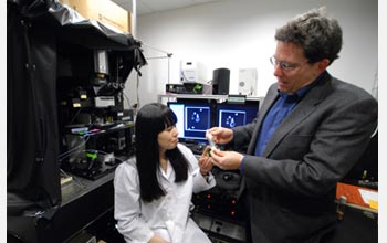
Biologist David Anderson and graduate student Suzuko Yorozu examine a vial containing fruit flies.
Biologist David Anderson and graduate student Suzuko Yorozu examine a vial containing fruit flies. In Anderson's lab researchers were able to cut a hole in the shell that covers the fly's brain for a detailed view. Having used sophisticated techniques to selectively visualize the activity of particular genes in the fly, the researchers could see when any neurons in the fly's brain were activated by a particular stimulus.
Credit: Bob Paz/Caltech
Images credited to the National Science Foundation, a federal agency, are in the public domain. The images were created by employees of the United States Government as part of their official duties or prepared by contractors as "works for hire" for NSF. You may freely use NSF-credited images and, at your discretion, credit NSF with a "Courtesy: National Science Foundation" notation.
Additional information about general usage can be found in Conditions.
Also Available:
Download the high-resolution JPG version of the image. (2.9 MB)
Use your mouse to right-click (Mac users may need to Ctrl-click) the link above and choose the option that will save the file or target to your computer.
Related story: Fruit Flies' Response to Wind Offers New Window to Neural Circuits