
Multimedia Gallery
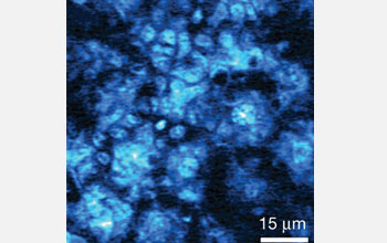
Image of the delivery of "toluidine blue O" to the deepest layer of skin in a mouse ear.
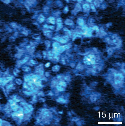
Stimulated emission image of the delivery of "toluidine blue O" to the same area of freshly cut mouse ear skin as in the second image. Image was taken at a depth of 25 micrometers 30 minutes after topical application of the molecular dye. A micrometer is one-millionth of a meter.
The image shows a rich distribution of toluidine blue O, or TBO, following the subcellular cytoplasm of basal keratinocytes. Basal keratinocytes are the major cellular constituents of skin and located in the deepest layer of skin.
The top and middle images support that the hydrophilic, or water loving, path is the main pathway for transdermal drug delivery of TBO. Subcellular localization of TBO is crucial because it influences both the level and the kinetics of inducing cellular death or apoptosis.
Credit: Wei Min and Sijia Lu, Department of Chemistry and Chemical Biology, Harvard University
Images credited to the National Science Foundation, a federal agency, are in the public domain. The images were created by employees of the United States Government as part of their official duties or prepared by contractors as "works for hire" for NSF. You may freely use NSF-credited images and, at your discretion, credit NSF with a "Courtesy: National Science Foundation" notation.
Additional information about general usage can be found in Conditions.
Also Available:
Download the high-resolution JPG version of the image. (150 KB)
Use your mouse to right-click (Mac users may need to Ctrl-click) the link above and choose the option that will save the file or target to your computer.
Related story: Seeing Previously Invisible Molecules for the First Time