
Multimedia Gallery
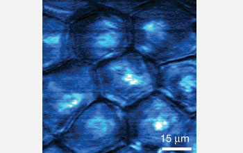
Image of the delivery of "toluidine blue O" to the outer most layer of skin in a mouse ear
Stimulated emission image of the delivery of "toluidine blue O" to an area of freshly cut mouse ear skin at the depth of three micrometers. The image was taken 30 minutes after topical application of toluidine blue O. A micrometer is one-millionth of a meter. Toluidine blue O, or TBO, is a molecular dye with an affinity for cancer cells in living organisms. The non-fluorescent drug is used in cancer therapy to locate cancer cells, which are then killed by radiation.
The image was taken at the surface layer of stratum corneum, which is the outer most layer of skin. The image shows TBO accumulated in the protein phase of the polygonal cells rather than in the lipid-rich intercellular space.
Credit: Wei Min and Sijia Lu, Department of Chemistry and Chemical Biology, Harvard University
Images credited to the National Science Foundation, a federal agency, are in the public domain. The images were created by employees of the United States Government as part of their official duties or prepared by contractors as "works for hire" for NSF. You may freely use NSF-credited images and, at your discretion, credit NSF with a "Courtesy: National Science Foundation" notation.
Additional information about general usage can be found in Conditions.
Also Available:
Download the high-resolution JPG version of the image. (144 KB)
Use your mouse to right-click (Mac users may need to Ctrl-click) the link above and choose the option that will save the file or target to your computer.
Related story: Seeing Previously Invisible Molecules for the First Time