
Multimedia Gallery
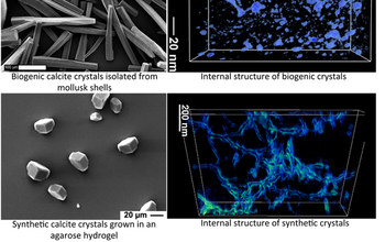
These images show calcite crystals (prisms).
Top left: Scanning electron micrograph (SEM) of calcite crystals (also called prisms) isolated from the prismatic layer of Atrina rigida (pen-shell mollusk).
Top right: 3-D reconstruction of the internal structure of the calcite prisms. The blue areas represent organic-filled voids within the crystal. The technique used to acquire this image is annular dark field scanning transmission electron microscopy (ADF-STEM) tomography.
Bottom left: SEM of synthetic calcite crystals grown in an agarose hydrogel.
Bottom right: ADF-STEM tomography showing a 3-D reconstruction of the internal structure of the synethic calcite crystals grown in an agarose hydrogel. The structures outlined in blue represent the agarose fibers that are incorporated into the single crystal of calcite.
Credit: Estroff Lab, Cornell University
Images credited to the National Science Foundation, a federal agency, are in the public domain. The images were created by employees of the United States Government as part of their official duties or prepared by contractors as "works for hire" for NSF. You may freely use NSF-credited images and, at your discretion, credit NSF with a "Courtesy: National Science Foundation" notation.
Additional information about general usage can be found in Conditions.
Also Available:
Download the high-resolution JPG version of the image. (1023.8 KB)
Use your mouse to right-click (Mac users may need to Ctrl-click) the link above and choose the option that will save the file or target to your computer.
Related story: Learning from biology to create new materials