
Multimedia Gallery
New technique could aid development of nanoscale biosensors
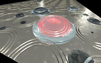
Plasmonic interferometers that have light emitters within them could make for better, more compact biosensors for a variety of applications.
More about this image
Researchers at Brown University’s School of Engineering have made an important fundamental advance that could make nanoscale plasmonic interferometry, a technique that combines nanotechnology with plasmonics -- the interaction between electrons in a metal and light, more practical. Plasmonic interferometry has the potential to enable compact, ultra-sensitive biosensors for a variety of applications.
The Brown team, led by Domenico Pacifici, a professor of engineering who oversaw the work with his postdoctoral researcher Dongfang Li and graduate student Jing Feng, has developed a technique that eliminates the need for highly specialized external light sources that deliver coherent light -- beams in which light waves are parallel, have the same wavelength and travel in-phase (meaning the peaks and valleys of the waves are aligned) -- which plasmonic interferometry normally requires. Without coherent light sources, the interferometers cannot produce usable interference patterns. But those kinds of light sources tend to be bulky, expensive, and require careful alignment and periodic recalibration to obtain a reliable optical response.
In the new method developed by Pacifici and his group, fluorescent light-emitting atoms are integrated directly within a tiny hole in the center of the interferometer. An external light source is still necessary to excite the internal emitters, but it doesn't have to be a specialized coherent source.
"This is a whole new concept for optical interferometry," said Pacifici, "an entirely new device."
"It has always been assumed that coherent light was necessary for plasmonic interferometry," adds Pacifici. "But we were able to disprove that assumption." The advance could enable more versatile and more compact devices.
This research was supported by National Science Foundation (NSF) grant CBET 11-59255.
To learn more about this research, see the NSF News From the Field story Advance could aid development of nanoscale biosensors. (Date image taken: 2016; date originally posted to NSF Multimedia Gallery: July 12, 2016)
Credit: Pacific Lab/Brown University
See other images like this on your iPhone or iPad download NSF Science Zone on the Apple App Store.
Images and other media in the National Science Foundation Multimedia Gallery are available for use in print and electronic material by NSF employees, members of the media, university staff, teachers and the general public. All media in the gallery are intended for personal, educational and nonprofit/non-commercial use only.
Images credited to the National Science Foundation, a federal agency, are in the public domain. The images were created by employees of the United States Government as part of their official duties or prepared by contractors as "works for hire" for NSF. You may freely use NSF-credited images and, at your discretion, credit NSF with a "Courtesy: National Science Foundation" notation.
Additional information about general usage can be found in Conditions.
Also Available:
Download the high-resolution JPG version of the image. (682.5 KB)
Use your mouse to right-click (Mac users may need to Ctrl-click) the link above and choose the option that will save the file or target to your computer.