
All Images
News Release 08-218
Next Generation Microscopy: No Stain, Big Gain
Researchers can monitor drug distribution and perform medical diagnostics rapidly using a new 3D imaging technique
This material is available primarily for archival purposes. Telephone numbers or other contact information may be out of date; please see current contact information at media contacts.

This figure shows SRS images for diffusion of retinoic acid (blue) and DMSO (green) through the top layer of the skin, or stratum corneum, which is the main barrier against topically applied drugs. Retinoic acid, a common drug for acne, diffuses through the lipids of the stratum corneum (polygonal shape), and is visualized by tuning the Raman shift into the characteristic band at 1570cm-1 (blue). DMSO, often used as a diffusion enhancer, goes deeper into the skin. It is hydrophilic (water-loving) so it avoids the lipid structures that retinoic acid diffuses through readily. This is highlighted using two-color SRS imaging tuned into the characteristic vibration of DMSO at 670cm-1 (green) and the vibration of typical skin lipids at 2845cm-1 (red) at a depth of ~65µm into the skin.
Credit: Image Courtesy of Chris Freudiger, Wei Min, Brain Saar, Harvard University, in collaboration with Pfizer's Jason Tsai
Download the high-resolution JPG version of the image. (177 KB)
Use your mouse to right-click (Mac users may need to Ctrl-click) the link above and choose the option that will save the file or target to your computer.

The researchers' findings appear in the December 19, 2008, issue of Science magazine.
Credit: Copyright 2008 AAAS
Download the high-resolution JPG version of the image. (312 KB)
Use your mouse to right-click (Mac users may need to Ctrl-click) the link above and choose the option that will save the file or target to your computer.
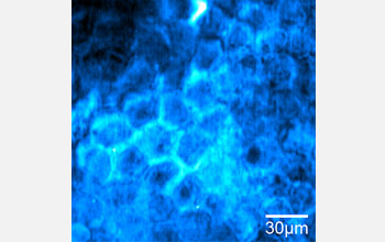
This SRS image shows diffusion of retinoic acid (blue), a common drug for acne, through the top layer of the skin, or stratum corneum. Retinoic acid diffuses through the lipids of the stratum corneum, seen here at the edges of skin cells (polygonal shape), and is visualized by tuning the Raman shift into the characteristic band at 1570cm-1.
Credit: Image Courtesy of Chris Freudiger, Wei Min, Brain Saar, Harvard University, in collaboration with Pfizer's Jason Tsai
Download the high-resolution JPG version of the image. (63 KB)
Use your mouse to right-click (Mac users may need to Ctrl-click) the link above and choose the option that will save the file or target to your computer.
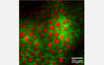
Two-color SRS imaging tuned into the characteristic vibration of DMSO at 670cm-1 (green) and the vibration of typical skin lipids at 2845cm-1 (red) at a depth of ~65µm into the skin. DMSO, often used as a diffusion enhancer, is hydrophilic (water-loving) so it avoids the lipid structures that retinoic acid diffuses through readily.
Credit: Image Courtesy of Chris Freudiger, Wei Min, Brain Saar, Harvard University, in collaboration with Pfizer's Jason Tsai
Download the high-resolution JPG version of the image. (51 KB)
Use your mouse to right-click (Mac users may need to Ctrl-click) the link above and choose the option that will save the file or target to your computer.