
All Images
News Release 11-047
Researchers Selectively Control Anxiety Pathways in the Brain
Study uses NSF-supported technology to identify neuronal circuitry
This material is available primarily for archival purposes. Telephone numbers or other contact information may be out of date; please see current contact information at media contacts.
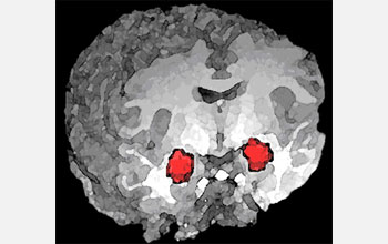
A new study supports the role of a brain region called the amygdala in processing anxiety. In this 3-D magnetic resonance imaging (MRI) rendering of a human brain, functional MRI (fMRI) activation of the amygdala is highlighted in red.
Credit: NIMH Clinical Brain Disorders Branch
Download the high-resolution JPG version of the image. (118 KB)
Use your mouse to right-click (Mac users may need to Ctrl-click) the link above and choose the option that will save the file or target to your computer.