
All Images
Research News
What Lies Beneath? Understanding Art Using Science

Top: Scenes From the Life of Saint John the Baptist (Francesco Granacci; ca. 1506-1507), egg tempera, oil, and gold on wood; 77.6×151.1 cm.
Bottom: Cross section of paint layers from Scenes from the Life of Saint John the Baptist, 20x objective, DIC light.
Credit: Please contact the copyright holder before reproducing.
Top: The Metropolitan Museum of Art. Purchase of Gwynne Andrews, Harris Brisbane Dick, Dodge, Fletcher, and Rogers; funds from various donors; gift from Ella Morris de Peyster and Mrs. Donald Oenslager; and gifts in memory of Robert Lehman, 1970 (1970.134.1) Image © The Metropolitan Museum of Art.
Bottom: Image © The Metropolitan Museum of Art, Department of Scientific Research.
Download the high-resolution JPG version of the image. (58 KB)
Use your mouse to right-click (Mac users may need to Ctrl-click) the link above and choose the option that will save the file or target to your computer.
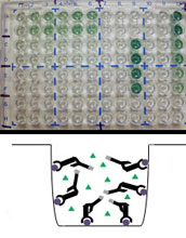
Top: ELISA plate after the enzyme-catalyzed color reaction. The colored product indicates the presence of a protein or gum of interest to researchers. Bottom: Schematic diagram of a positive reaction in one well of the ELISA plate.
Credit: Please contact the copyright holder before reproducing. Image © The Metropolitan Museum of Art, Department of Scientific Research
Download the high-resolution JPG version of the image. (47 KB)
Use your mouse to right-click (Mac users may need to Ctrl-click) the link above and choose the option that will save the file or target to your computer.
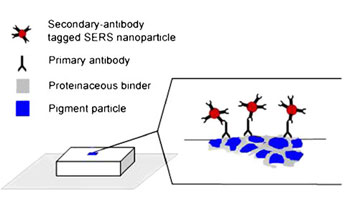
Schematic diagram of Indirect ImmunoSERS Assay.
Credit: Please contact the copyright holder before reproducing. Image © The Metropolitan Museum of Art, Department of Scientific Research.
Download the high-resolution JPG version of the image. (40 KB)
Use your mouse to right-click (Mac users may need to Ctrl-click) the link above and choose the option that will save the file or target to your computer.
Julie Arslanoglu, Associate Research Scientist, Department of Scientific Research, Metropolitan Museum of Art.
Credit: Please contact the copyright holder before reproducing. Image © The Metropolitan Museum of Art, Department of Scientific Research.
Download the high-resolution JPG version of the image. (1.3 MB)
Use your mouse to right-click (Mac users may need to Ctrl-click) the link above and choose the option that will save the file or target to your computer.

John Loike, Ph.D., Co-Director of Graduate Studies Department Physiology & Cellular Biophysics, Columbia University, College of Physicians and Surgeons.
Credit: Please contact the copyright holder before reproducing. Image © Columbia University, College of Physicians and Surgeons.
Download the high-resolution JPG version of the image. (21 KB)
Use your mouse to right-click (Mac users may need to Ctrl-click) the link above and choose the option that will save the file or target to your computer.

Dina Georgas (left) and Stephanie Zaleski (right), Barnard College undergraduates, interns of the Department of Scientific Research at the Metropolitan Museum of Art.
Credit: Please contact the copyright holder before reproducing. Image © The Metropolitan Museum of Art, Department of Scientific Research.
Download the high-resolution JPG version of the image. (1.4 MB)
Use your mouse to right-click (Mac users may need to Ctrl-click) the link above and choose the option that will save the file or target to your computer.