Shining Light on the Nanoscale
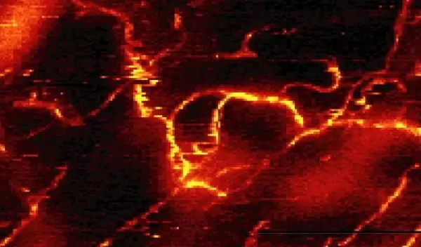
In 2003 researchers created the highest-resolution optical image up to that point, revealing structures as small as carbon nanotubes just a few billionths of an inch across.
The new method, developed by scientists at the University of Rochester, with colleagues from Portland State University and Harvard University, should literally shed light on previously inaccessible chemical and structural information in samples as small as the proteins in a cell's membrane.
"This is the highest-resolution optical spectroscopic measurement ever made," said Lukas Novotny, professor of optics at Rochester. "There are other methods that can see smaller structures, but none use light, which is rich in information." The research, which appeared in the March 7, 2003, issue of Physical Review Letters, was supported by NSF.
The Rochester team's light-based technique, called near-field Raman microscopy, allows researchers to glean a great deal of visual information. Other ultra-high-resolution imaging techniques, such as atomic force microscopes, detect the presence of objects and image them but do not have the ability to directly view the light bouncing off an object.
To light up the nanoscale, Novotny and Hartschuh sharpen a gold wire to a point just a few billionths of an inch across. A laser then shines against the side of the gold tip, creating a tiny bubble of electromagnetic energy that interacts with the vibrations of the atoms in the sample. This interaction, called Raman scattering, releases packets of light from the sample that can be used to identify the chemical composition of the material.
With the new technique, Novotny and his colleague Achim Hartschuh can determine a material's composition as well as its structure. Novotny and his team are also eager to learn characteristics of newly discovered structures, such as crossed or interconnected carbon nanotubes.
The ultimate vision for the project is to garner the information light provides from the proteins on a membrane, which would open the door to designer medicines that could kill harmful cells, repair damaged cells or even identify never-before-seen strains of disease.
In about two years, Novotny and Hartschuh think they will be able to refine the system, already with a resolution of 20 nanometers (billionths of a meter), so that they can image proteins, which are only 5 to 20 nanometers wide, with eventual resolutions revealing much smaller molecules.
-- David Hart


