
Multimedia Gallery
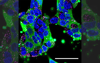
Confocal microscopy image of human embryonic kidney cells
Confocal microscopy image of human embryonic kidney cells. Nanoparticles filled with human immunoglobulin were delivered to the cells. The cell nucleus is blue, endosomes (which sequester materials taken up by cells) are labeled in pink, and the immunoglobulin proteins are labeled with a green fluorescent tag. The nanoparticle's cargo has spread widely through most of the cells (green) without it being trapped in endosomes.
[Research supported by National Science Foundation Graduate Research Fellowships DGE 0707427 and DGE 1232825]
Learn more about this research in the Johns Hopkins news story Little size holds big impact: Johns Hopkins scientists develop nanocontainer to ship titan-size gene therapies and drugs into cells. (Date image taken: unknown; date originally posted to NSF Multimedia Gallery: March 24, 2020)
Credit: Yuan Rui, Johns Hopkins Medicine,Johns Hopkins University
Images and other media in the National Science Foundation Multimedia Gallery are available for use in print and electronic material by NSF employees, members of the media, university staff, teachers and the general public. All media in the gallery are intended for personal, educational and nonprofit/non-commercial use only.
Images credited to the National Science Foundation, a federal agency, are in the public domain. The images were created by employees of the United States Government as part of their official duties or prepared by contractors as "works for hire" for NSF. You may freely use NSF-credited images and, at your discretion, credit NSF with a "Courtesy: National Science Foundation" notation.
Additional information about general usage can be found in Conditions.
Also Available:
Download the high-resolution JPG version of the image. (3.0 MB)
Use your mouse to right-click (Mac users may need to Ctrl-click) the link above and choose the option that will save the file or target to your computer.