
Multimedia Gallery
 All images in this series
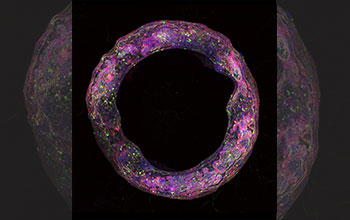
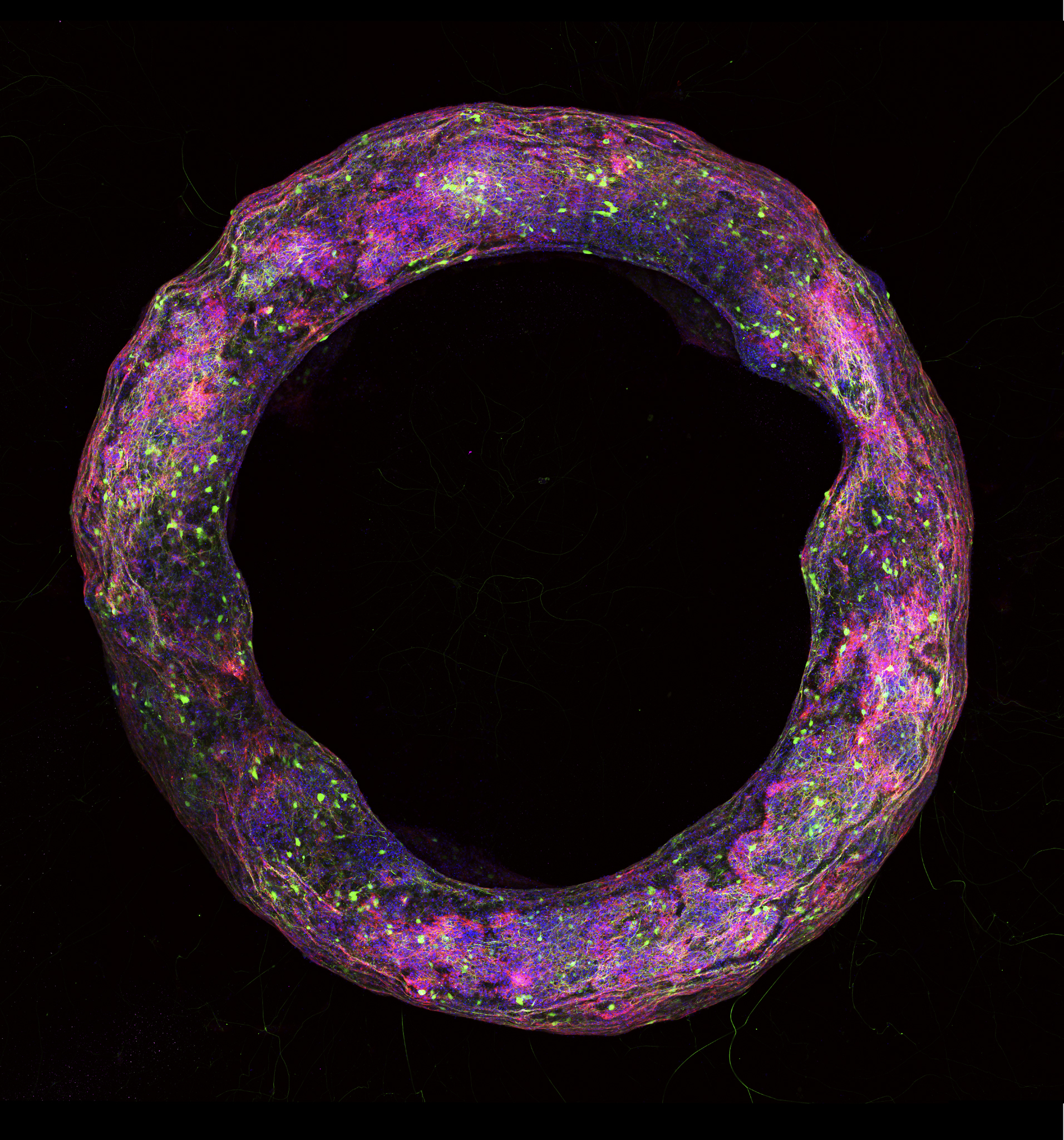
Toroid neural tissue geometry
This neural tissue geometry in the shape of a toroid was formed with a new biofabrication method developed by researchers at the University of Illinois and imaged using confocal microscopy.
[Research supported by National Science Foundation grants DGE 1144245, DGE 1735252 and CBET 3939511.]
Learn more in the University of Illinois news story Illinois team develops first of a kind in-vitro 3D neural tissue model. (Date image taken: 2019; date originally posted to NSF Multimedia Gallery: June 8, 2020)
Credit: Image courtesy of Gelson J Pagan-Diaz; Institute of Genomic Biology, University of Illinois at Urbana Champaign
Images and other media in the National Science Foundation Multimedia Gallery are available for use in print and electronic material by NSF employees, members of the media, university staff, teachers and the general public. All media in the gallery are intended for personal, educational and nonprofit/non-commercial use only.
Images credited to the National Science Foundation, a federal agency, are in the public domain. The images were created by employees of the United States Government as part of their official duties or prepared by contractors as "works for hire" for NSF. You may freely use NSF-credited images and, at your discretion, credit NSF with a "Courtesy: National Science Foundation" notation.
Additional information about general usage can be found in Conditions.
Also Available:
Download the high-resolution JPG version of the image. (3.0 MB)
Use your mouse to right-click (Mac users may need to Ctrl-click) the link above and choose the option that will save the file or target to your computer.