
Multimedia Gallery
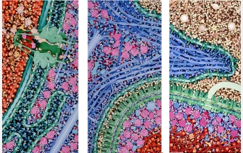
Macrophage and Bacterium
This series of three paintings, titled "Macrophage and Bacterium 2,000,000X," shows a macrophage engulfing a bacterium. Macrophages circulate through the blood, searching for bacterial infection. When bacteria are found, macrophages engulf and digest them. Only a portion of the two cells, where a pseudopod of the macrophage is extending over the bacterium, is shown. The original paintings are 1 meter tall. At this magnification, the macrophage would fill most of a building.
These paintings, by David S. Goodsell of the Scripps Research Institute in La Jolla, Calif., are on display at the Center for Integrative Molecular Biosciences at Scripps. They include all of the macromolecules in the two cells and in the surrounding blood serum. The small organic molecules and water, which fill all the space between the macromolecules, are omitted. In the paintings, the cell membranes and their associated proteins are colored green, the cytoplasm is colored blue and purple, and the nuclear material is colored red and orange. The blood serum proteins are in yellow and brown.
The illustration took second place in the second annual "Science and Engineering Visualization Challenge" competition. The international contest is designed to recognize outstanding achievements by scientists, engineers, visualization specialists and artists in the use of visual media to promote understanding of research results and scientific phenomena. The competition is sponsored jointly by the National Science Foundation and the journal Science, published by the American Association for the Advancement of Science (AAAS). Further information about the competition is available at http://www.nsf.gov/od/lpa/events/sevc/start.cfm.
More about this Image
For the past decade, David S. Goodsell has created watercolor paintings that reveal the molecular world inside cells. Images of this world are not accessible by experimental means--microscopy reveals the ultrastructure of cells and molecular biology reveals the atomic structure of molecules--but the intermediate level, where molecules combine to create living cells, is largely invisible. Goodsell's paintings synthesize information from both ends of this experimental spectrum, showing portions of cells at molecular detail.
"Macrophage and Bacterium 2,000,000X" depicts a cross section through a macrophage in the process of engulfing a bacterium. The illustration shows only a portion of the two cells in the area of contact and the individual macromolecules involved in immune recognition, as well as the macromolecules involved in the basic processes of life. The colors highlight the different functional regions of the cells: In the macrophage, the nucleus is shown in red and orange, the cytoplasm is shown in blue and purple, and membranes and the endoplasmic reticulum are shown in greens. The bacterium is colored similarly to highlight the similarities between the two cells and the blood is shown in browns and yellows at upper right.
Goodsell creates these paintings as tools for exploration. Viewers can start by identifying major structures, like the large nuclear pore at left, or the bacterial flagellum and motor at right, and then move on to find other familiar structures. Included in the painting are RNA polymerase and DNA polymerase in action, many ribosomes, antibodies, action filaments, a microtubule with kinesin and hundreds of other biological molecules. (Year of image: 2002)
Credit: David S. Goodsell, Scripps Research Institute
See other images like this on your iPhone or iPad download NSF Science Zone on the Apple App Store.
Special Restrictions: This image is unrestricted for private and educational use only. Permission is required from the artist for re-publication/reproduction or commercial use. Contact David S. Goodsell via e-mail at goodsell@scripps.edu.
Images and other media in the National Science Foundation Multimedia Gallery are available for use in print and electronic material by NSF employees, members of the media, university staff, teachers and the general public. All media in the gallery are intended for personal, educational and nonprofit/non-commercial use only.
Images credited to the National Science Foundation, a federal agency, are in the public domain. The images were created by employees of the United States Government as part of their official duties or prepared by contractors as "works for hire" for NSF. You may freely use NSF-credited images and, at your discretion, credit NSF with a "Courtesy: National Science Foundation" notation.
Additional information about general usage can be found in Conditions.
Also Available:
Download the high-resolution JPG version of the image. (7 MB)
Use your mouse to right-click (Mac users may need to Ctrl-click) the link above and choose the option that will save the file or target to your computer.