
Multimedia Gallery
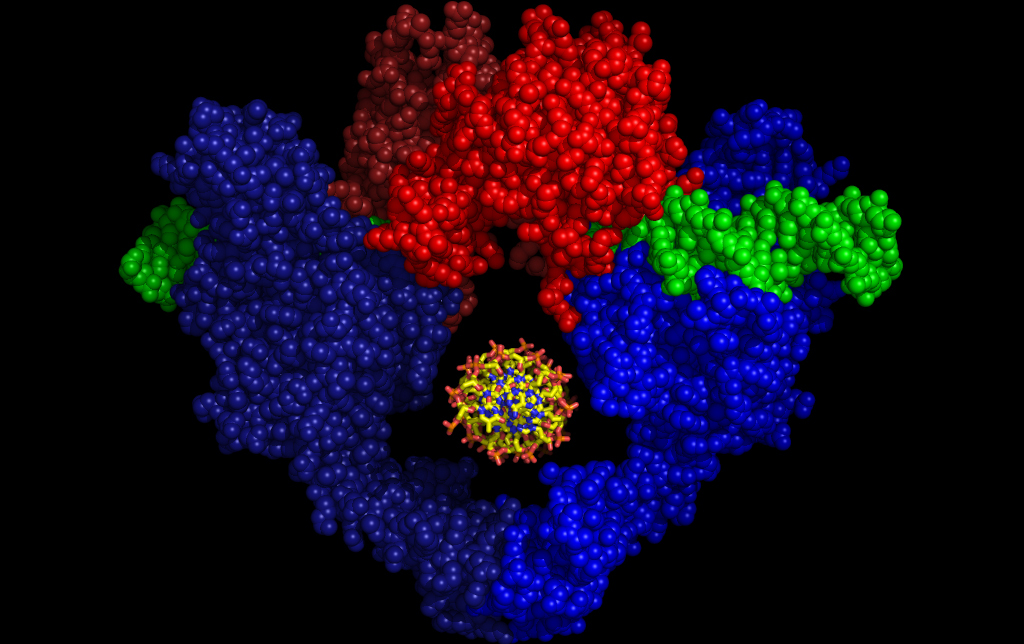
DNA Binding and Cleavage Fragment
DNA binding and cleavage fragment of yeast topoisomerase II.
Model for the DNA binding and cleavage fragment of yeast topoisomerase II (blue/red spheres) bound to a separated DNA strand (green spheres). One protomer is shaded darker than the other to highlight the protein's two subunits. A second DNA (viewed end-on, yellow sticks) is modeled into a large internal hole present in the enzyme. This figure represents a likely conformational and substrate-bound intermediate of this region of the topoisomerase during its duplex DNA passage reaction. This research was performed in the lab of James Berger, professor of biochemistry and molecular biology at the University of California, Berkeley.
Research in Berger's lab focuses on understanding the molecular basis of protein assembly and function. In particular, we aim to develop mechanistic models that explain how proteins physically exploit shape and chemistry to form large complexes and transduce energy into work. The primary focus of my group's investigations is on proteins that replicate, manipulate, and organize nucleic acids in the cell, and on metabolic enzymes required for maintaining nucleic acids in the pathogenic bacterium, M. tuberculosis. A combination of structural analyses, such as X-ray crystallography, small-angle X-ray scattering and EM, coupled with biphysical and biochemical experimentation, forms the core of our methodological approach. (Date of Image: 1995)
Credit: Figure courtesy James M. Berger, UC-Berkeley
See other images like this on your iPhone or iPad download NSF Science Zone on the Apple App Store.
Images and other media in the National Science Foundation Multimedia Gallery are available for use in print and electronic material by NSF employees, members of the media, university staff, teachers and the general public. All media in the gallery are intended for personal, educational and nonprofit/non-commercial use only.
Images credited to the National Science Foundation, a federal agency, are in the public domain. The images were created by employees of the United States Government as part of their official duties or prepared by contractors as "works for hire" for NSF. You may freely use NSF-credited images and, at your discretion, credit NSF with a "Courtesy: National Science Foundation" notation.
Additional information about general usage can be found in Conditions.
Also Available:
Download the high-resolution JPG version of the image. (402 KB)
Use your mouse to right-click (Mac users may need to Ctrl-click) the link above and choose the option that will save the file or target to your computer.