
Multimedia Gallery
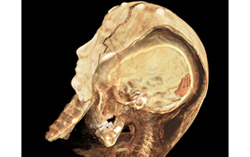
"An Egyptian Child Mummy"
"An Egyptian Child Mummy"
A high-resolution computed tomography (CT) scan of an Egyptian child mummy. For 75 years, this child mummy resided in the Rosicrucian Egyptian Museum in San Jose, California, its body unseen by human eyes, its story a mystery. In 2005, a team of researchers joined computer engineer Paul Brown of Stanford University School of Medicine, in unraveling the threads of this mystery using the latest in imaging technology -- an AXIOM Siemens scanner, one of only five CT scanners in the world capable of producing such high-resolution images.
Stanford University radiology's state-of-the-art scanner generated 2D slices as thin as 200 microns -- several times thinner than the 750 micron slices used to create the popular 3D visualization of King Tutankhamen's mummy. In fact, at 92 gigabytes, Stanford radiology's child mummy scans generated nearly 35 times more information than the scans conducted on King Tut.
Equipped with the most detailed 3D models ever created of a mummy, the team of experts reconstructed 60,000 exceptionally high-resolution 2D scans to help them give life to this mummy without disturbing its delicate form. The result is the highest quality interactive visualization of a mummy ever seen. Analysis of the data revealed that the 2,000-year-old mummy is the remains of a 4 to 5-year-old girl who, researchers concluded, likely died unexpectedly from an infectious disease.
This photo was awarded first place in the photography category of the 2006 Science & Engineering Visualization Challenge competition, sponsored by the National Science Foundation and the Journal Science. The competition is held each year to recognize outstanding achievements by scientists, engineers, visualization specialists and artists who are innovators in using visual media to promote the understanding of research results and scientific phenomena. To learn more about the competition and view all the winning entries, see the Science & Engineering Visualization Challenge Special Report. (Date of Image: June 2005)
Credit: W. Paul Brown, DDS, principal investigator
Images and other media in the National Science Foundation Multimedia Gallery are available for use in print and electronic material by NSF employees, members of the media, university staff, teachers and the general public. All media in the gallery are intended for personal, educational and nonprofit/non-commercial use only.
Images credited to the National Science Foundation, a federal agency, are in the public domain. The images were created by employees of the United States Government as part of their official duties or prepared by contractors as "works for hire" for NSF. You may freely use NSF-credited images and, at your discretion, credit NSF with a "Courtesy: National Science Foundation" notation.
Additional information about general usage can be found in Conditions.
Also Available:
Download the high-resolution TIF version of the image. (16.9 MB)
Use your mouse to right-click (Mac users may need to Ctrl-click) the link above and choose the option that will save the file or target to your computer.