
Multimedia Gallery
Pathogenic Fungus Candida albicans (Image 1)
Pathogenic Fungus Candida albicans (Image 1)
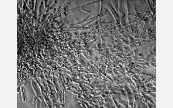
The pathogenic fungus Candida albicans, grown on a solid agar medium obtained with a light microscope. Some cells have assumed a small, round form, while others are long, filamentous hyphal cells. The ability to shift forms is thought to be crucial for invasion of--and survival within--the different tissues that Candida infects.
Quinn Mitrovich, a postdoctoral scientist at the University of California, San Francisco, is studying what allows Candida to enter the bloodstream and bodily environs with the hope of discovering ways to block these molecular events and halt raging Candida infections. To learn more about this research, see the UCSF Today story, Learning Cut-and-Paste Rules to Fight a Deadly Fungus. (Date of Image: Aug. 14, 2006) [One of three related images. See Next Image.]
Credit: Dr. Quinn Mitrovich, University of California, San Francisco
Images and other media in the National Science Foundation Multimedia Gallery are available for use in print and electronic material by NSF employees, members of the media, university staff, teachers and the general public. All media in the gallery are intended for personal, educational and nonprofit/non-commercial use only.
Images credited to the National Science Foundation, a federal agency, are in the public domain. The images were created by employees of the United States Government as part of their official duties or prepared by contractors as "works for hire" for NSF. You may freely use NSF-credited images and, at your discretion, credit NSF with a "Courtesy: National Science Foundation" notation.
Additional information about general usage can be found in Conditions.
Also Available:
Download the high-resolution JPG version of the image. (178 KB)
Use your mouse to right-click (Mac users may need to Ctrl-click) the link above and choose the option that will save the file or target to your computer.