
Multimedia Gallery
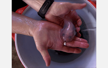
Antifreeze fish
Paraliparis Devriesi, named after researcher Arthur DeVries of the University of Illinois, an American biologist who has studied Antarctic fish for 40 years.
More about this image
Arthur DeVries' research focuses on proteins developed by Antarctic fish that act as antifreeze, allowing them to live in the frigid, freezing waters of the Southern Ocean.
These antifreeze proteins are a group of unique macromolecules that help some polar and subpolar marine bony fish avoid freezing in their icy habitats. The proteins were discovered by DeVries from fish that he collected at McMurdo Station in Antarctica while he was a graduate student at Stanford University in the early 1960s.
The waters of the Southern Ocean are so cold that temperate and tropical fish would freeze if they were placed in this environment. The presence of salt in seawater allows it to remain liquid until about -1.9 degrees Celsius, almost 2 degrees below the freezing temperature of fresh water. The antifreeze proteins -- along with normal body salts -- depress the freezing point of blood and body fluids to 2.5 degrees Celsius, slightly below the freezing point of seawater. These proteins bind to and inhibit growth of ice crystals within body fluids through an absorption inhibition process. The proteins attach to small ice crystals, stemming their growth. This mechanism that inhibits further growth of the ice crystals remains under study, but apparently Antarctic fish are able to survive with very small ice crystals present in their body fluids.
Researchers have found similar compounds in other cold-water fish, insects, plants, fungi and bacteria. Because of the numerous potential benefits of protecting tissue from damage by freezing, private companies have begun to explore the use of these compounds in increasing freeze tolerance of commercial plants, improving farm fish production in cold climates, extending shelf life of frozen foods, improving cryosurgery (i.e., surgery involving the freezing of certain tissues) and improving preservation of tissues for transplant or transfusion in medicine. (Date of Image: Nov. 16, 2002)
Credit: Melanie Conner, NSF
Images and other media in the National Science Foundation Multimedia Gallery are available for use in print and electronic material by NSF employees, members of the media, university staff, teachers and the general public. All media in the gallery are intended for personal, educational and nonprofit/non-commercial use only.
Images credited to the National Science Foundation, a federal agency, are in the public domain. The images were created by employees of the United States Government as part of their official duties or prepared by contractors as "works for hire" for NSF. You may freely use NSF-credited images and, at your discretion, credit NSF with a "Courtesy: National Science Foundation" notation.
Additional information about general usage can be found in Conditions.
Also Available:
Download the high-resolution JPG version of the image. (248 KB)
Use your mouse to right-click (Mac users may need to Ctrl-click) the link above and choose the option that will save the file or target to your computer.