
Multimedia Gallery
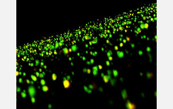
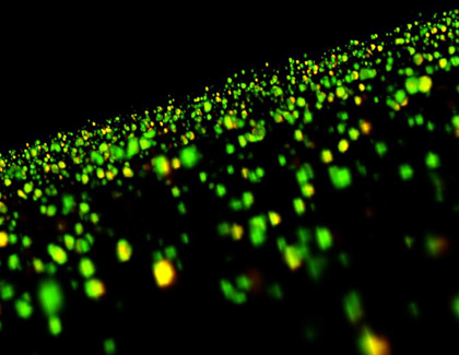
First-Ever Image of Brain Changes
First-Ever Image of Brain Changes
Image of brain dendritic spines after long-term potentiation.
Researchers from the University of California, Irvine, developed the first images to show the actual physical changes in brain cells that are thought to underlie memory, a discovery that is already uncovering clues about memory loss linked to cognitive disorders. To learn more about this research, see the UC-Irvine press release "UC Irvine Researchers Reveal First Images of Brain Changes Associated with Memory". (Date of Image: March 2007)
Credit: Lulu Y. Chen, Christopher S. Rex, Christine M. Gall, and Gary Lynch, University of California, Irvine.
Images and other media in the National Science Foundation Multimedia Gallery are available for use in print and electronic material by NSF employees, members of the media, university staff, teachers and the general public. All media in the gallery are intended for personal, educational and nonprofit/non-commercial use only.
Images credited to the National Science Foundation, a federal agency, are in the public domain. The images were created by employees of the United States Government as part of their official duties or prepared by contractors as "works for hire" for NSF. You may freely use NSF-credited images and, at your discretion, credit NSF with a "Courtesy: National Science Foundation" notation.
Additional information about general usage can be found in Conditions.
Also Available:
Download the high-resolution JPG version of the image. (98 KB)
Use your mouse to right-click (Mac users may need to Ctrl-click) the link above and choose the option that will save the file or target to your computer.